
What you will learn in this chapter
- Describe the structure of a nucleotide and the Watson–Crick double helix model of DNA.
- Explain the experiments that proved DNA is the genetic material (Griffith; Avery, MacLeod and McCarty; Hershey–Chase).
- Explain semiconservative replication using the Meselson–Stahl experiment, and leading vs lagging strands.
- Describe transcription in prokaryotes and eukaryotes, including splicing, capping and tailing.
- State the salient features of the genetic code and explain translation and the lac operon.
- Explain the goals and salient features of the Human Genome Project and the steps of DNA fingerprinting.
📘 Open this chapter on NCERT official website (ncert.nic.in) · The same NCERT chapter is taught in both CBSE and UP Board.
Molecular Basis of Inheritance — Notes
🖼️ Diagrams are now right inside the notes. Tap any picture to zoom and see the name and job of each part; 🧊 3D opens the 3D model.डायग्राम अब नोट्स के अंदर ही हैं। किसी भी चित्र पर टैप करें — ज़ूम करें और हर भाग का नाम व कार्य देखें; 🧊 3D से 3D मॉडल खुलता है।Diagrams ab notes ke andar hi hain. Kisi bhi picture par tap kijiye — zoom kijiye aur har part ka naam aur kaam dekhiye; 🧊 3D se 3D model khulta hai.
Revise these first (prerequisites)
- Basic idea of cell, nucleus and chromosome
- Proteins are made of amino acids (Class 11 Biomolecules)
- Chapter 4: concept of gene and allele
- Simple meaning of a hydrogen bond (Chemistry)
DNA = the instruction book of life
Inside every cell, DNA is like a recipe book that states which protein is to be made and when. A gene is one 'chapter' of this book. Proteins do most of the work in the body, so the instructions written in DNA determine our traits.
Building block of DNA — the nucleotide
DNA is made of small units called nucleotides: one nitrogenous base + one sugar (deoxyribose) + one phosphate. There are four bases: A, T, G, C. It is like writing an entire book using an alphabet of just 4 letters.
DNA double helix (Watson–Crick model)डीएनए द्विकुंडलिनी (वॉटसन–क्रिक मॉडल)DNA double helix (Watson–Crick model)NCERT Fig 5.3Two antiparallel strands, sugar–phosphate backbone outside, base pairs inside: A=T (2 H-bonds), G≡C (3). One turn = 3.4 nm, 10 bp.दो प्रतिसमांतर रज्जुक, बाहर शर्करा–फॉस्फेट, अंदर क्षारक युग्म: A=T (2 H-बंध), G≡C (3)। एक घुमाव = 3.4 nm, 10 bp।2 antiparallel strands, bahar sugar–phosphate, andar base pairs: A=T (2 H-bonds), G≡C (3). Ek turn = 3.4 nm, 10 bp. Double helix — a twisted ladder
DNA is made of two strands that look like a twisted ladder. The 'steps' of the ladder are pairs of bases: A always pairs with T and G always pairs with C. This model was given by Watson and Crick in 1953.
DNA double helix (Watson–Crick model)डीएनए द्विकुंडलिनी (वॉटसन–क्रिक मॉडल)DNA double helix (Watson–Crick model)NCERT Fig 5.3Two antiparallel strands, sugar–phosphate backbone outside, base pairs inside: A=T (2 H-bonds), G≡C (3). One turn = 3.4 nm, 10 bp.दो प्रतिसमांतर रज्जुक, बाहर शर्करा–फॉस्फेट, अंदर क्षारक युग्म: A=T (2 H-बंध), G≡C (3)। एक घुमाव = 3.4 nm, 10 bp।2 antiparallel strands, bahar sugar–phosphate, andar base pairs: A=T (2 H-bonds), G≡C (3). Ek turn = 3.4 nm, 10 bp.🧊 See in 3D: DNA double helix3D में देखें: DNA द्विकुंडली3D dekhiye: DNA double helix Replication = making a copy
Before a cell divides, DNA makes an exact copy of itself — replication (प्रतिकृतियन). Both strands unwind and each old strand acts as a template for a new strand. So every new DNA molecule has one old and one new strand (semiconservative).
Replicating forkप्रतिकृति द्विशाख (रेप्लिकेशन फ़ोर्क)Replicating forkNCERT Fig 5.8At the fork the leading strand is made continuously, the lagging strand in short Okazaki fragments joined by DNA ligase.फोर्क पर अग्रग रज्जुक लगातार बनता है, पश्चग रज्जुक छोटे ओकाज़ाकी खंडों में, जिन्हें DNA लाइगेज़ जोड़ता है।Fork par leading strand lagatar banta hai, lagging strand chhote Okazaki fragments mein, jinhe DNA ligase jodta hai. DNA → RNA → Protein
The information in DNA is first copied into mRNA (transcription / अनुलेखन), then the code of the mRNA is read to make protein (translation / स्थानांतरण). This flow is called the central dogma. Think of it this way: the master recipe (DNA) stays safe, and only its photocopy (mRNA) goes to the kitchen.
Transcription unit, transcription and RNA processingअनुलेखन इकाई, अनुलेखन और आरएनए संसाधनTranscription unit, transcription and RNA processingNCERT Fig 5.9–5.11Promoter → structural gene → terminator. In eukaryotes the hnRNA is capped, tailed and spliced (introns out) into mRNA.प्रवर्तक → संरचनात्मक जीन → समापक। यूकैरियोट में hnRNA पर कैप, पूँछ और स्प्लाइसिंग (इंट्रॉन बाहर) होकर mRNA बनता है।Promoter → structural gene → terminator. Eukaryotes mein hnRNA par capping, tailing aur splicing (introns bahar) hokar mRNA banta hai. Translation on the ribosomeराइबोसोम पर अनुवादनTranslation on the ribosomeNCERT Fig 5.13The ribosome moves along mRNA codon by codon; tRNAs bring amino acids and peptide bonds join them.राइबोसोम mRNA पर कोडॉन दर कोडॉन चलता है; tRNA अमीनो अम्ल लाते हैं और पेप्टाइड बंध उन्हें जोड़ते हैं।Ribosome mRNA par codon by codon chalta hai; tRNA amino acids laate hain aur peptide bonds unhe jodte hain. Genetic code — 3-letter words
mRNA is read in groups of 3 bases — each group is a codon that specifies one amino acid. Reading starts at AUG (start) and stops at UAA, UAG or UGA (stop).
tRNA — the adapter moleculetRNA — अनुकूलक अणुtRNA — the adapter moleculeNCERT Fig 5.12Clover-leaf adapter: anticodon loop reads the mRNA codon, the 3′ end (amino acid acceptor) carries the amino acid.तिपतिया पत्ती जैसा अनुकूलक: प्रतिकोडॉन लूप mRNA कोडॉन पढ़ता है, 3′ सिरा (अमीनो अम्ल ग्राही) अमीनो अम्ल लाता है।Clover-leaf adapter: anticodon loop mRNA codon padhta hai, 3′ end (amino acid acceptor) amino acid laata hai.
Quick check ✔
1. In DNA, Adenine (A) pairs with which base?
2. What is the process of forming mRNA from DNA called?
Basics clear? Now move on to Learn in Detail 👆
As per NCERT 2026-27 — every point you need for the board exam.
Nucleotide
Nitrogenous base + pentose sugar = nucleoside; + phosphate = nucleotide. Purines: A, G. Pyrimidines: C, T (DNA), U (RNA). Nucleotides are joined by 3'-5' phosphodiester bonds; the base and sugar are joined by an N-glycosidic bond.
DNA double helix (Watson–Crick model)डीएनए द्विकुंडलिनी (वॉटसन–क्रिक मॉडल)DNA double helix (Watson–Crick model)NCERT Fig 5.3Two antiparallel strands, sugar–phosphate backbone outside, base pairs inside: A=T (2 H-bonds), G≡C (3). One turn = 3.4 nm, 10 bp.दो प्रतिसमांतर रज्जुक, बाहर शर्करा–फॉस्फेट, अंदर क्षारक युग्म: A=T (2 H-बंध), G≡C (3)। एक घुमाव = 3.4 nm, 10 bp।2 antiparallel strands, bahar sugar–phosphate, andar base pairs: A=T (2 H-bonds), G≡C (3). Ek turn = 3.4 nm, 10 bp. DNA double helix
Watson & Crick (1953), based on X-ray data of Wilkins and Rosalind Franklin. Two antiparallel strands; A=T (2 H-bonds), G≡C (3 H-bonds). Chargaff's rule: A = T, G = C. Pitch 3.4 nm, ~10 bp per turn, 0.34 nm between bp.
DNA double helix (Watson–Crick model)डीएनए द्विकुंडलिनी (वॉटसन–क्रिक मॉडल)DNA double helix (Watson–Crick model)NCERT Fig 5.3Two antiparallel strands, sugar–phosphate backbone outside, base pairs inside: A=T (2 H-bonds), G≡C (3). One turn = 3.4 nm, 10 bp.दो प्रतिसमांतर रज्जुक, बाहर शर्करा–फॉस्फेट, अंदर क्षारक युग्म: A=T (2 H-बंध), G≡C (3)। एक घुमाव = 3.4 nm, 10 bp।2 antiparallel strands, bahar sugar–phosphate, andar base pairs: A=T (2 H-bonds), G≡C (3). Ek turn = 3.4 nm, 10 bp.🧊 See in 3D: DNA double helix3D में देखें: DNA द्विकुंडली3D dekhiye: DNA double helix Length of DNA
A human diploid cell has ~6.6 × 10⁹ bp → ~2.2 m of DNA. E. coli: 4.6 × 10⁶ bp. Lambda phage: 48502 bp.
DNA packaging
Prokaryotes: nucleoid. Eukaryotes: an octamer of positively charged histones (lysine, arginine) + DNA = nucleosome (~200 bp). Beads-on-string → chromatin fibre. Euchromatin = loose, active; heterochromatin = dense, inactive.
Nucleosome and beads-on-stringन्यूक्लियोसोम और धागे पर मोतीNucleosome and beads-on-stringNCERT Fig 5.4DNA (~200 bp) wraps around a histone octamer = nucleosome; a chain of nucleosomes looks like beads-on-string.DNA (~200 bp) हिस्टोन अष्टक पर लिपटता है = न्यूक्लियोसोम; न्यूक्लियोसोम की शृंखला धागे पर मोतियों जैसी।DNA (~200 bp) histone octamer par lipat-ta hai = nucleosome; nucleosomes ki chain beads-on-string jaisi.🧊 See in 3D: DNA + nucleosome3D में देखें: DNA + न्यूक्लियोसोम3D dekhiye: DNA + nucleosome Discovery of the genetic material
Griffith (1928): Streptococcus pneumoniae (S & R strains) → transforming principle. Avery, MacLeod, McCarty: it is DNA. Hershey–Chase (1952): bacteriophage, radioactive ³²P (DNA) entered, ³⁵S (protein) did not → DNA is the genetic material.
Griffith's transformation experiment (1928)ग्रिफिथ का रूपांतरण प्रयोग (1928)Griffith's transformation experiment (1928)S strain kills, R strain doesn't, heat-killed S doesn't — but heat-killed S + live R kills: R was 'transformed'.S प्रभेद मारता है, R नहीं, ताप-मृत S नहीं — पर ताप-मृत S + जीवित R मारता है: R 'रूपांतरित' हुआ।S strain maarta hai, R nahi, heat-killed S nahi — par heat-killed S + live R maarta hai: R 'transform' ho gaya. Hershey–Chase experiment (1952)हर्शे–चेज़ प्रयोग (1952)Hershey–Chase experiment (1952)NCERT Fig 5.5Radioactive P (in DNA) entered the bacteria, radioactive S (in protein) stayed outside — so DNA is the genetic material.रेडियोधर्मी P (DNA में) जीवाणु में गया, रेडियोधर्मी S (प्रोटीन में) बाहर रहा — अतः DNA आनुवंशिक पदार्थ है।Radioactive P (DNA mein) bacteria ke andar gaya, radioactive S (protein mein) bahar raha — isliye DNA genetic material hai. RNA world
The first genetic material was RNA. RNA also acts as a catalyst (ribozyme), but due to its 2'-OH group it is reactive and unstable. That is why the more stable DNA became the genetic material.
Replication
Semiconservative — Meselson & Stahl (1958), using ¹⁵N in E. coli. Enzyme: DNA-dependent DNA polymerase (5'→3'); begins at the origin of replication, forming a replication fork. Leading strand is continuous, lagging strand is discontinuous (Okazaki fragments, joined by DNA ligase). Occurs in the S-phase.
Meselson and Stahl's experiment (1958)मेसेल्सन और स्टाल का प्रयोग (1958)Meselson and Stahl's experiment (1958)NCERT Fig 5.7¹⁵N DNA grown in ¹⁴N: after 1 generation all hybrid (intermediate band), after 2 generations half hybrid, half light → semiconservative.¹⁵N DNA को ¹⁴N में: 1 पीढ़ी बाद सब संकर (बीच की पट्टी), 2 पीढ़ी बाद आधा संकर, आधा हल्का → अर्धसंरक्षी।¹⁵N DNA ko ¹⁴N mein: 1 generation baad sab hybrid (beech ki band), 2 generation baad aadha hybrid, aadha light → semiconservative. Replicating forkप्रतिकृति द्विशाख (रेप्लिकेशन फ़ोर्क)Replicating forkNCERT Fig 5.8At the fork the leading strand is made continuously, the lagging strand in short Okazaki fragments joined by DNA ligase.फोर्क पर अग्रग रज्जुक लगातार बनता है, पश्चग रज्जुक छोटे ओकाज़ाकी खंडों में, जिन्हें DNA लाइगेज़ जोड़ता है।Fork par leading strand lagatar banta hai, lagging strand chhote Okazaki fragments mein, jinhe DNA ligase jodta hai. Central dogma & transcription
DNA → RNA → protein (Crick). Transcription unit: promoter, structural gene, terminator. Template strand 3'→5', coding strand 5'→3' (same sequence as mRNA, except T in place of U). Bacteria: a single RNA polymerase, σ factor (initiation), ρ factor (termination).
Transcription unit, transcription and RNA processingअनुलेखन इकाई, अनुलेखन और आरएनए संसाधनTranscription unit, transcription and RNA processingNCERT Fig 5.9–5.11Promoter → structural gene → terminator. In eukaryotes the hnRNA is capped, tailed and spliced (introns out) into mRNA.प्रवर्तक → संरचनात्मक जीन → समापक। यूकैरियोट में hnRNA पर कैप, पूँछ और स्प्लाइसिंग (इंट्रॉन बाहर) होकर mRNA बनता है।Promoter → structural gene → terminator. Eukaryotes mein hnRNA par capping, tailing aur splicing (introns bahar) hokar mRNA banta hai. Eukaryotic transcription
RNA pol I → rRNA (28S, 18S, 5.8S); pol II → hnRNA (precursor of mRNA); pol III → tRNA, 5S rRNA, snRNA. hnRNA undergoes splicing (introns removed, exons joined), capping (methyl guanosine triphosphate added at the 5' end) and tailing (poly-A added at the 3' end).
Transcription unit, transcription and RNA processingअनुलेखन इकाई, अनुलेखन और आरएनए संसाधनTranscription unit, transcription and RNA processingNCERT Fig 5.9–5.11Promoter → structural gene → terminator. In eukaryotes the hnRNA is capped, tailed and spliced (introns out) into mRNA.प्रवर्तक → संरचनात्मक जीन → समापक। यूकैरियोट में hnRNA पर कैप, पूँछ और स्प्लाइसिंग (इंट्रॉन बाहर) होकर mRNA बनता है।Promoter → structural gene → terminator. Eukaryotes mein hnRNA par capping, tailing aur splicing (introns bahar) hokar mRNA banta hai. Genetic code
Triplet code; 64 codons: 61 for amino acids, 3 stop (UAA, UAG, UGA). AUG = start + methionine. Unambiguous, degenerate, comma-less, nearly universal. George Gamow suggested the triplet code; Nirenberg and Khorana deciphered it.
tRNA & translation
tRNA = adapter molecule (clover-leaf / inverted L); has an anticodon loop and an amino acid acceptor end. Charging (aminoacylation) → initiation (AUG) → elongation (peptide bond; 23S rRNA of ribosome = ribozyme) → termination (release factor). UTRs on both sides.
tRNA — the adapter moleculetRNA — अनुकूलक अणुtRNA — the adapter moleculeNCERT Fig 5.12Clover-leaf adapter: anticodon loop reads the mRNA codon, the 3′ end (amino acid acceptor) carries the amino acid.तिपतिया पत्ती जैसा अनुकूलक: प्रतिकोडॉन लूप mRNA कोडॉन पढ़ता है, 3′ सिरा (अमीनो अम्ल ग्राही) अमीनो अम्ल लाता है।Clover-leaf adapter: anticodon loop mRNA codon padhta hai, 3′ end (amino acid acceptor) amino acid laata hai. Translation on the ribosomeराइबोसोम पर अनुवादनTranslation on the ribosomeNCERT Fig 5.13The ribosome moves along mRNA codon by codon; tRNAs bring amino acids and peptide bonds join them.राइबोसोम mRNA पर कोडॉन दर कोडॉन चलता है; tRNA अमीनो अम्ल लाते हैं और पेप्टाइड बंध उन्हें जोड़ते हैं।Ribosome mRNA par codon by codon chalta hai; tRNA amino acids laate hain aur peptide bonds unhe jodte hain. Lac operon (Jacob & Monod)
Structural genes: z (β-galactosidase), y (permease), a (transacetylase); i gene = repressor. Lactose (inducer) inactivates the repressor → transcription ON. This is negative regulation.
The lac operonलैक ओपेरॉनThe lac operonNCERT Fig 5.14No lactose: repressor blocks the operator (genes OFF). Lactose (inducer) binds the repressor → genes z, y, a switch ON.लैक्टोज़ नहीं: दमनकारी प्रचालक को रोकता है (जीन बंद)। लैक्टोज़ (प्रेरक) दमनकारी से जुड़ता है → z, y, a जीन चालू।Lactose nahi: repressor operator ko block karta hai (genes OFF). Lactose (inducer) repressor se judta hai → z, y, a genes ON. HGP & DNA fingerprinting
Human Genome Project (1990–2003): ~3 × 10⁹ bp; about 30,000 genes estimated; 99.9% bases are the same in all humans; chromosome 1 has the most genes (2968), Y the fewest (231); less than 2% of the genome codes for proteins; ~1.4 million SNPs. DNA fingerprinting (Alec Jeffreys): VNTR / satellite DNA probe, Southern blotting → forensics, paternity test.
DNA fingerprinting (VNTR)डीएनए अंगुलिछापी (VNTR)DNA fingerprinting (VNTR)NCERT Fig 5.16VNTRs differ in copy number between people, so the band pattern on the gel is unique for each person.VNTR की प्रति-संख्या हर व्यक्ति में अलग, इसलिए जेल पर पट्टियों का पैटर्न हर व्यक्ति का अलग।VNTRs ki copy number har insaan mein alag, isliye gel par band pattern har vyakti ka unique.
The concept in depth — the why and the how. Everything stays within the NCERT syllabus.
Why is DNA better than RNA as genetic material?
Every nucleotide of RNA has a 2'-OH group, which makes it reactive and easily degradable; DNA lacks it, so DNA is more stable. Thymine (in place of uracil) also gives DNA extra stability. Being reactive, RNA also mutates quickly — RNA viruses evolve fast. Hence DNA is better for stable storage, and RNA is better for fast expression/catalysis — and the first genetic material is believed to have been RNA (RNA world).
Why is the lagging strand discontinuous?
DNA polymerase can add nucleotides only in the 5'→3' direction, and the two strands are antiparallel. Therefore, synthesis is continuous on one strand (template 3'→5'), and discontinuous on the other in small pieces (Okazaki fragments), which are later joined by DNA ligase. Replication starts only at the origin — this fact is used in the cloning vectors (ori) of Chapter 9.
Replicating forkप्रतिकृति द्विशाख (रेप्लिकेशन फ़ोर्क)Replicating forkNCERT Fig 5.8At the fork the leading strand is made continuously, the lagging strand in short Okazaki fragments joined by DNA ligase.फोर्क पर अग्रग रज्जुक लगातार बनता है, पश्चग रज्जुक छोटे ओकाज़ाकी खंडों में, जिन्हें DNA लाइगेज़ जोड़ता है।Fork par leading strand lagatar banta hai, lagging strand chhote Okazaki fragments mein, jinhe DNA ligase jodta hai. The logic of the Meselson–Stahl experiment
E. coli was first grown in 15N (heavy) medium, then shifted to 14N; DNA was separated by CsCl density gradient centrifugation. After 20 minutes (one generation), all the DNA was hybrid (intermediate); after 40 minutes, hybrid and light DNA were present in equal amounts. If replication were conservative, separate heavy and light bands would have appeared in the very first generation — so this is proof of semiconservative replication.
Meselson and Stahl's experiment (1958)मेसेल्सन और स्टाल का प्रयोग (1958)Meselson and Stahl's experiment (1958)NCERT Fig 5.7¹⁵N DNA grown in ¹⁴N: after 1 generation all hybrid (intermediate band), after 2 generations half hybrid, half light → semiconservative.¹⁵N DNA को ¹⁴N में: 1 पीढ़ी बाद सब संकर (बीच की पट्टी), 2 पीढ़ी बाद आधा संकर, आधा हल्का → अर्धसंरक्षी।¹⁵N DNA ko ¹⁴N mein: 1 generation baad sab hybrid (beech ki band), 2 generation baad aadha hybrid, aadha light → semiconservative. Extra steps of eukaryotic transcription
Eukaryotes have three RNA polymerases: I (rRNAs 28S, 18S, 5.8S), II (hnRNA → mRNA), III (tRNA, 5S rRNA, snRNA). hnRNA contains introns, which are removed by splicing; a methyl guanosine triphosphate cap is added at the 5' end (capping) and 200–300 adenylate residues are added at the 3' end (tailing). Split genes (exons + introns) are probably an 'ancient' feature and a reminder of the RNA world.
Transcription unit, transcription and RNA processingअनुलेखन इकाई, अनुलेखन और आरएनए संसाधनTranscription unit, transcription and RNA processingNCERT Fig 5.9–5.11Promoter → structural gene → terminator. In eukaryotes the hnRNA is capped, tailed and spliced (introns out) into mRNA.प्रवर्तक → संरचनात्मक जीन → समापक। यूकैरियोट में hnRNA पर कैप, पूँछ और स्प्लाइसिंग (इंट्रॉन बाहर) होकर mRNA बनता है।Promoter → structural gene → terminator. Eukaryotes mein hnRNA par capping, tailing aur splicing (introns bahar) hokar mRNA banta hai. Lac operon — genes switched ON/OFF as needed
In the lac operon, the z (β-galactosidase), y (permease) and a (transacetylase) genes are transcribed together (polycistronic). The repressor made by the regulator gene i binds to the operator and blocks transcription. When lactose (inducer) is present, it binds to the repressor and inactivates it, and the genes are switched ON. This is negative regulation — the cell saves energy because the enzyme is made only when lactose is present.
The lac operonलैक ओपेरॉनThe lac operonNCERT Fig 5.14No lactose: repressor blocks the operator (genes OFF). Lactose (inducer) binds the repressor → genes z, y, a switch ON.लैक्टोज़ नहीं: दमनकारी प्रचालक को रोकता है (जीन बंद)। लैक्टोज़ (प्रेरक) दमनकारी से जुड़ता है → z, y, a जीन चालू।Lactose nahi: repressor operator ko block karta hai (genes OFF). Lactose (inducer) repressor se judta hai → z, y, a genes ON. Basis of DNA fingerprinting: polymorphism
99.9% of the DNA of any two humans is identical; the differences lie in repetitive DNA. The copy number of VNTRs (satellite DNA) differs from person to person — they are detected by Southern blot/hybridisation using a probe. This pattern is inherited from the parents, so it is useful in paternity disputes and forensic cases. The technique was developed by Alec Jeffreys.
DNA fingerprinting (VNTR)डीएनए अंगुलिछापी (VNTR)DNA fingerprinting (VNTR)NCERT Fig 5.16VNTRs differ in copy number between people, so the band pattern on the gel is unique for each person.VNTR की प्रति-संख्या हर व्यक्ति में अलग, इसलिए जेल पर पट्टियों का पैटर्न हर व्यक्ति का अलग।VNTRs ki copy number har insaan mein alag, isliye gel par band pattern har vyakti ka unique.
Common confusions — cleared up
Template strand vs Coding strand
RNA is made from the template strand (3'→5'). The sequence of the coding strand (5'→3') is the same as the RNA (with U in place of T) — that is why the sequence is always written as that of the coding strand.
Degenerate vs Ambiguous code
Degenerate = several codons for one amino acid (correct). Ambiguous = one codon for several amino acids — the genetic code is unambiguous; one codon specifies only one amino acid.
Nucleoside vs Nucleotide
Nucleoside = base + sugar. Nucleotide = base + sugar + phosphate.
Euchromatin vs Heterochromatin
Euchromatin is loosely packed, stains light and is transcriptionally active. Heterochromatin is densely packed, stains dark and is inactive.
Nucleosome and beads-on-stringन्यूक्लियोसोम और धागे पर मोतीNucleosome and beads-on-stringNCERT Fig 5.4DNA (~200 bp) wraps around a histone octamer = nucleosome; a chain of nucleosomes looks like beads-on-string.DNA (~200 bp) हिस्टोन अष्टक पर लिपटता है = न्यूक्लियोसोम; न्यूक्लियोसोम की शृंखला धागे पर मोतियों जैसी।DNA (~200 bp) histone octamer par lipat-ta hai = nucleosome; nucleosomes ki chain beads-on-string jaisi.
Video
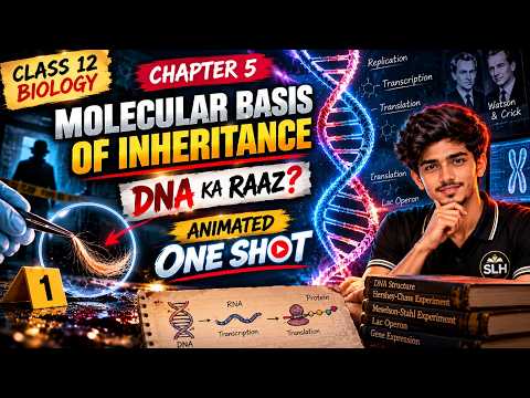
▶ Molecular Basis of Inheritance Class 12 One Shot | NCERT Biology Chapter 5 | Board Exam 2027 · 58 min
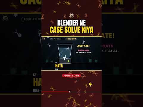
1-minute revision Shorts



Diagrams (tap & zoom)
All 11 diagrams of this chapter — tap one to open itइस अध्याय के सभी 11 डायग्राम — खोलने के लिए टैप करेंIs chapter ke saare 11 diagrams — kholne ke liye tap kijiye
 DNA double helix (Watson–Crick model)डीएनए द्विकुंडलिनी (वॉटसन–क्रिक मॉडल)Fig 5.3
 Nucleosome and beads-on-stringन्यूक्लियोसोम और धागे पर मोतीFig 5.4
 Griffith's transformation experiment (1928)ग्रिफिथ का रूपांतरण प्रयोग (1928)
 Hershey–Chase experiment (1952)हर्शे–चेज़ प्रयोग (1952)Fig 5.5
 Meselson and Stahl's experiment (1958)मेसेल्सन और स्टाल का प्रयोग (1958)Fig 5.7
 Replicating forkप्रतिकृति द्विशाख (रेप्लिकेशन फ़ोर्क)Fig 5.8
 Transcription unit, transcription and RNA processingअनुलेखन इकाई, अनुलेखन और आरएनए संसाधनFig 5.9–5.11
 tRNA — the adapter moleculetRNA — अनुकूलक अणुFig 5.12
 Translation on the ribosomeराइबोसोम पर अनुवादनFig 5.13
 The lac operonलैक ओपेरॉनFig 5.14
 DNA fingerprinting (VNTR)डीएनए अंगुलिछापी (VNTR)Fig 5.16
Detail: Watson–Crick model: two antiparallel polynucleotide chains; bases face inward and are joined by H-bonds — A=T (2 bonds), G≡C (3 bonds); a purine (A, G) always pairs with a pyrimidine (T, C). The helix is right-handed, pitch 3.4 nm, ~10 bp per turn, 0.34 nm between adjacent bp.
Original SLH diagram · Ladder model of DNA: two antiparallel sugar-phosphate backbones (5′ to 3′ and 3′ to 5′), adenine pairs with thymine by two hydrogen bonds, guanine with cytosine by three.
Important questions with answers
Q1. What did the Hershey–Chase experiment prove?
Hershey and Chase (1952) labelled the DNA of a bacteriophage with radioactive phosphorus (³²P) and its protein with radioactive sulphur (³⁵S). After infection, only ³²P was found inside the bacteria, not ³⁵S. This proved that the viral DNA enters the bacteria — i.e., DNA, not protein, is the genetic material.
Q2. What is semiconservative replication?
In replication, both strands of DNA separate and act as templates. Each new DNA molecule has one old (parental) strand and one new strand. This was proved by Meselson and Stahl in 1958 through their ¹⁵N experiment.
Q3. What is the inducer in the lac operon?
Lactose (actually allolactose) is the inducer. It binds to the repressor protein and inactivates it, allowing RNA polymerase to transcribe the z, y and a genes.
Q4. What are the features of the genetic code?
The code is triplet (64 codons): 61 codons code for amino acids and 3 are stop codons; one codon specifies only one amino acid (unambiguous), but one amino acid can have several codons (degenerate); the code is read without commas, is nearly universal, and AUG acts as the start codon and also codes for methionine.
UP Board previous year questions (2022–2026)
These questions are from real UP Board Intermediate papers (copies from a third-party archive, read and verified by SLH). Also shown: in how many 2022–2026 papers (different sets) a question from this topic appeared. See year-wise papers →
- “Which of the following is known as universal codon? (A) AAA (B) CCC (C) GGG (D) UUU”
UP Board 20261 marksQuestions from this topic: 16 papers (2022, 2023, 2024, 2025, 2026) - “Which nitrogenous base is not present in DNA? (A) Thymine (B) Uracil (C) Adenine (D) Guanine”
UP Board 20261 marksQuestions from this topic: 14 papers (2022, 2023, 2024, 2025, 2026) - “Explain transfer RNA (tRNA) as an adaptor molecule.”
UP Board 20263 marksQuestions from this topic: 10 papers (2022, 2023, 2024, 2025, 2026) - “Name the two strands of the structural gene DNA of a transcription unit.”
UP Board 20262 marksQuestions from this topic: 8 papers (2022, 2023, 2025, 2026) - “What do you understand by DNA fingerprinting technique? Explain its process.”
UP Board 20265 marksQuestions from this topic: 8 papers (2022, 2023, 2024, 2026) - “Write the important goals of Human Genome Project (HGP).”
UP Board 20253 marksQuestions from this topic: 7 papers (2022, 2023, 2024, 2025) - “Describe the properties of genetic material.”
UP Board 20263 marksQuestions from this topic: 6 papers (2022, 2023, 2024, 2025, 2026)
Practice quiz
Tap an option — get instant right/wrong feedback and an explanation. Wrong answers are saved in "My mistakes".
1. How many hydrogen bonds are there between G and C in DNA?
2. Who proved semiconservative replication?
3. Which of these is a stop codon?
4. What does the 'z' gene of the lac operon code for?
5. In the Hershey–Chase experiment, what was DNA labelled with?
6. On which strand are Okazaki fragments formed?
7. Which RNA polymerase synthesises hnRNA in eukaryotes?
8. Who developed the technique of DNA fingerprinting?
Quick revision checklist
Before the exam — try explaining each topic aloud for 1 minute. If you get stuck, reread it in the notes above.
- Nucleotide
- DNA double helix
- Length of DNA
- DNA packaging
- Discovery of the genetic material
- RNA world
- Replication
- Central dogma & transcription
- Eukaryotic transcription
- Genetic code
- tRNA & translation
- Lac operon (Jacob & Monod)
- HGP & DNA fingerprinting